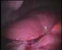
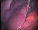
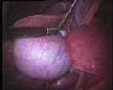
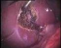
First, the surgeon creates holes and inserts his instruments. The operative photograph 1 shows the liver when the endoscope is introduced. The second action of the surgeon is to raise the right lobe of the liver so as to reach the gall bladder as show in photograph 2. Next the surgeon applies clips to and then cuts the cystic duct (photograph 3). Finally he uses a bipolar cautery instrument to coagulate the wound due to the ablation (photograph 4).
 |
 |
 |
 |
| photograph 1 | photograph 2 | photograph 3 | photograph 4 |