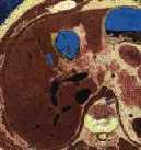
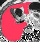
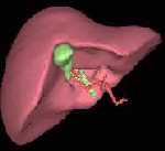
In order to have realistic simulated images, we work from NLM images. As show in the first picture, we take the slices corresponding to the liver. On each slice, we color and extract the liver points (picture 2) and create a volumetric model (picture 3). On this picture, one can see the liver and the gall bladder (in green). We also extract the vascular system of the liver showing in the animated image.
picture 1
picture 2
Model of the liver
Model of the Liver in transparency rendering with all the vascular system