-
CT-Scan Image Processing for the 3D Study of the Evolution of the Human Skull
/
Traitement d'images scanographiques appliqué à l'étude tridimensionnelle de l'évolution de la forme du crâne humain
-
| Authors / Auteurs |
Gérard Subsol (1)
Bertand Mafart (2)
Denis Méline (1)
Alain Silvestre (3)
Marie-Antoinette de Lumley (2)
|
-
| Authors References / References des auteurs |
(1) Projet EPIDAURE, Institut National de Recherche en Informatique et en Automatique, Sophia Antipolis
(2) Laboratoire d'Anthropologie, Faculté de Médecine de Marseille, Université de la Méditerranée
(3) Hôpital d'Instruction des Armées Laveran, Marseille
|
-
| Abstract | Resume |
A first algorithm extracts automatically the "crest lines" from the CT-Scan
images of the skull of a contemporary man (data provided by G. Quatrehomme,
University of Nice, France) and a prehistoric man, Broken-Hill (data provided
by D. Dean, Case Western Reserve University, Cleveland, USA). These lines
correspond to the most salient lines on the skull surface. They are used as
landmarks to find automatically the homologous points between the skulls. From
these matched points, we compute a volumetric transformation based on spline
functions that superimposes the two skulls. The transformation is then
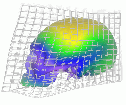
interpolated to create intermediate transformations. We extract a 3D surface
model of the skull of the contemporary man from the CT-Scan image and we apply
successively the intermediate transformations to visualise the evolution of the
skull shape between the prehistoric and contemporary man.
This work is currently extended to the study of other fossil skulls, in
particular, that of the Tautavel Man. We also plan to use 3D morphometry
methods based on crest lines to quantify the evolution of the skull shape and
to characterise various groups [1,3]. Lastly, we consider applications in
facial reconstruction [2] to synthesise the face of the prehistoric man based
on the skull shape.
|
Un premier algorithme extrait automatiquement des " lignes de crête " à
partir de scanographies de crânes d'un homme contemporain (données fournies
par G. Quatrehomme, Université de Nice) et préhistorique - homme de
Broken-Hill (données fournie par D. Dean, Université Case Western de
Cleveland - États-Unis). Ces lignes correspondent aux lignes saillantes de
la surface crânienne. Elles servent de repères à un algorithme de mise en
correspondance pour trouver automatiquement les points homologues entre les
deux crânes. À partir de ces points appariés, on calcule une transformation
de l'espace, fondée sur des fonctions splines, qui superpose les deux crânes.
Cette transformation est ensuite interpolée pour créer des transformations
intermédiaires. À partir de l'image scanographique, on extrait alors un
modèle surfacique du crâne de l'homme contemporain auquel on applique
successivement les transformations intermédiaires pour visualiser l'évolution
du crâne entre l'homme préhistorique et contemporain.
Ces travaux sont actuellement étendus à l'étude du crâne d'autres fossiles,
en particulier, l'homme de Tautavel. Nous prévoyons aussi d'utiliser des
méthodes de morphométrie tridimensionnelles qui se fondent sur les lignes
de crête pour quantifier l'évolution de la forme du crâne et caractériser
les différentes lignées [1,3]. Enfin, nous envisageons des applications en
reconstruction faciale [2] pour synthétiser le visage de l'homme préhistorique
à partir du crâne.
|
-
| References |
[1] D.Dean. The Middle Pleistocene Homo erectus/Homo sapiens Transition: New Evidence from Space Curve Statistics. Ph.D. thesis, The City University of New York, 1993.
[2] G. Quatrehomme, S. Cotin, G. Subsol, H. Delingette, Y. Garidel, G. Grevin, M. Fidrich, P. Bailet, A. Ollier. A Fully Three-Dimensional Method for Facial Reconstruction Based on Deformable Models. Journal of Forensic Sciences, 42(4):649-652, July 1997.
[3] G. Subsol. Construction automatique d'atlas anatomiques morphométriques à partir d'images médicales tridimensionnelles. Ph.D. thesis, École Centrale Paris, December 1995. In French. Electronic version: http://www.inria.fr/RRRT/TU-0379.html.
|
-
| Misc | Remarque |
Summited and accepted at ...... Creteil.
|
Soumis et accepter au colloque de Creteil.
|
-
| Electronic version | Version telechargeable |
Work in progress....
|
Travail en cours...
|
|


