![\fbox{\includegraphics[width=3cm]{olea006_050.eps}}](img33.gif) |
![\fbox{\includegraphics[width=3cm]{olea006_050.mask.eps}}](img34.gif) |
![\fbox{\includegraphics[width=3cm]{olea006_050.mask_inside.eps}}](img35.gif) |
![\fbox{\includegraphics[width=3cm]{olea006_050.mask_exine.eps}}](img36.gif) |
| Original image | Complete grain | Inside grain | Exine |
The characteristics are searched in their possible locations (complete grain, exine, or inside the grain).
![\fbox{\includegraphics[width=3cm]{olea006_050.eps}}](img33.gif) |
![\fbox{\includegraphics[width=3cm]{olea006_050.mask.eps}}](img34.gif) |
![\fbox{\includegraphics[width=3cm]{olea006_050.mask_inside.eps}}](img35.gif) |
![\fbox{\includegraphics[width=3cm]{olea006_050.mask_exine.eps}}](img36.gif) |
| Original image | Complete grain | Inside grain | Exine |
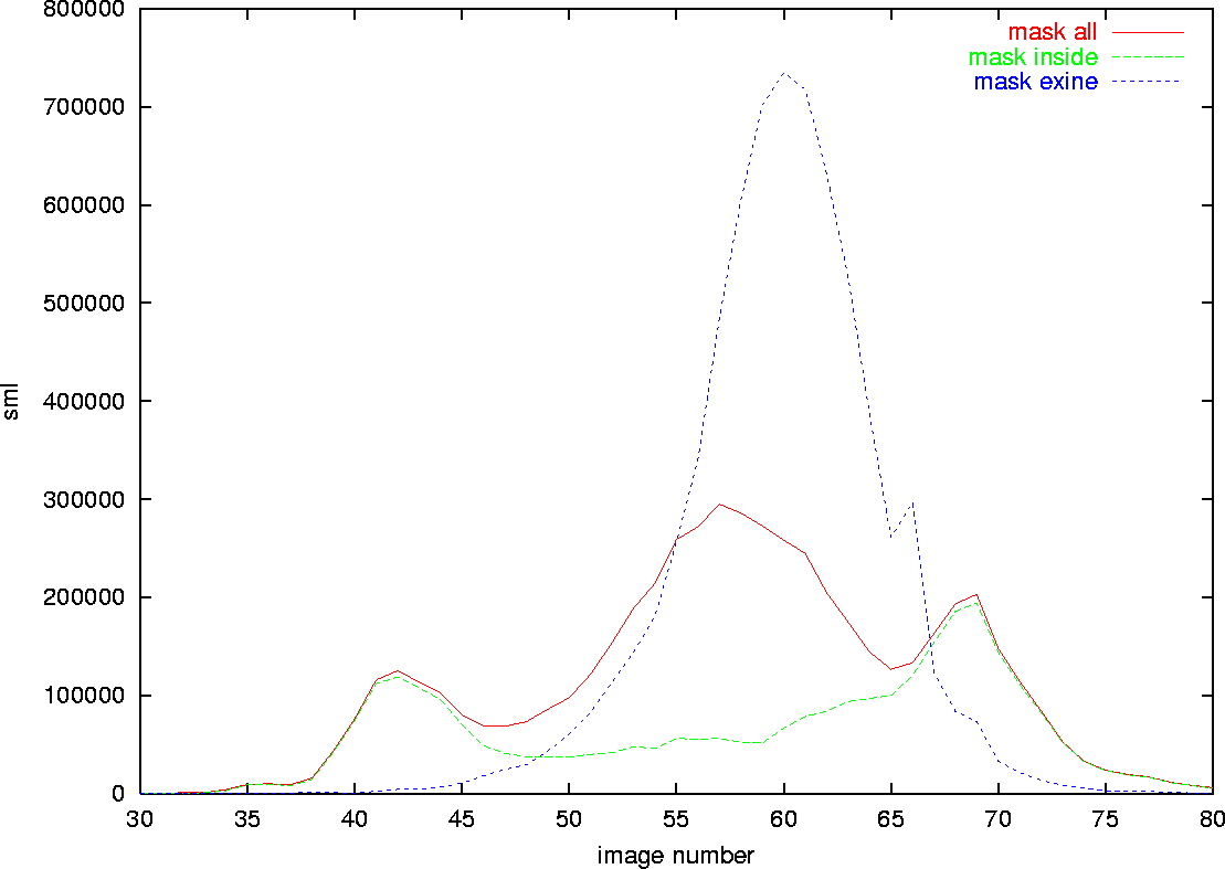
The image sequence can be analyzed to find the images of interest, i.e. the images that contains clear (on focus) characteristics.

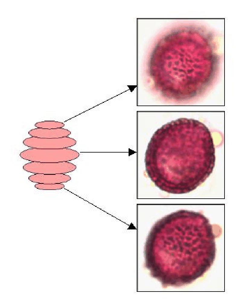
The pore of the Poaceae can appear anywhere on its surface, but is visible through several image.
![\fbox{\includegraphics[width=3cm]{poaceae002_035.eps}}](img38.gif) |
![\fbox{\includegraphics[width=3cm]{poaceae002_050.eps}}](img39.gif) |
![\fbox{\includegraphics[width=3cm]{poaceae002_065.eps}}](img40.gif) |
![\fbox{\includegraphics[width=3cm]{poaceae002_080.eps}}](img41.gif) |
| image 35/100 | image 50/100 | image 65/100 | image 80/100 |
![\fbox{\includegraphics[width=3cm]{poaceae002_035.pore.eps}}](img42.gif) |
![\fbox{\includegraphics[width=3cm]{poaceae002_050.pore.eps}}](img43.gif) |
![\fbox{\includegraphics[width=3cm]{poaceae002_065.pore.eps}}](img44.gif) |
![\fbox{\includegraphics[width=3cm]{poaceae002_080.pore.eps}}](img45.gif) |
The cytoplasm of the Cupressaceae is located in the center of the grain. It is known to have a star-like shape, but this cannot be verifier for many examples. It appears mostly as a bright zone visible in several images near the center.
![\fbox{\includegraphics[width=3cm]{cupressaceae003_040.eps}}](img46.gif) |
![\fbox{\includegraphics[width=3cm]{cupressaceae003_050.eps}}](img47.gif) |
![\fbox{\includegraphics[width=3cm]{cupressaceae003_060.eps}}](img48.gif) |
| image 40/100 | image 50/100 | image 60/100 |
![\fbox{\includegraphics[width=3cm]{cupressaceae003_040.cytoplasm.eps}}](img49.gif) |
![\fbox{\includegraphics[width=3cm]{cupressaceae003_050.cytoplasm.eps}}](img50.gif) |
![\fbox{\includegraphics[width=3cm]{cupressaceae003_060.cytoplasm.eps}}](img51.gif) |
The reticulum is located on the grain surface, but can be analyzed mostly on the top of bottom image of the grain (which show the best view fo the surface).

Not all pollen types are reticulated. So the steps to follow are:
![\fbox{\includegraphics[width=3cm]{olea001_035.eps}}](img53.gif) |
![\fbox{\includegraphics[width=3cm]{olea001_035.olea.eps}}](img54.gif) |
![\fbox{\includegraphics[width=3cm]{olea001_035.oleaperimeter.eps}}](img55.gif) |
![\fbox{\includegraphics[width=3cm]{olea001_035.oleamin3.eps}}](img56.gif) |