Large scale simulator of biological retina
INRIA CeCILL C open-source license (2007)
Partially supported by the EC IP project FP6-015879 FACETS
Neuromathcomp research-team, INRIA Sophia Antipolis - Méditerranée
 |
Virtual
Retina Large scale simulator of biological retina INRIA CeCILL C open-source license (2007) Partially supported by the EC IP project FP6-015879 FACETS Neuromathcomp research-team, INRIA Sophia Antipolis - Méditerranée |
| Overview |
Web-service | Results |
Customization |
Download | Publications | Contact |
 |
| Output
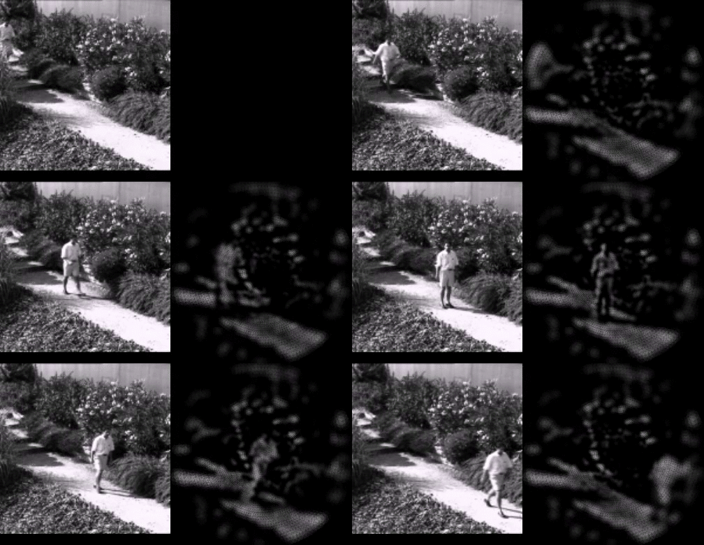
of the software What you will get from Virtual Retina is a list of spikes in an ASCII file, as decribed in the sample result below.
The number on the left is the index of the spiking cell, the number on the right is the corresponding time of spike, in seconds after the beginning of the simulation. Reconstruction In order to visualize the results, Virtual Retina also offers you the possibility to reconstruct a sequence, where each emitted spike adds a spot to the image, for a duration of 50 ms, and with a size that depends on the distance from the center of the retina (far from center means less cells, but associated to bigger spots in the reconstruction).  Spike emission of a
modeled
channel of ON parvocellular
cells,
in response to five seconds of video input. This reconstruction procedure allows to observe the qualitative effect of the contrast gain control stage. We give below two examples of retinal output on the same input image (left) , with (right) and without (middle) a contrast-gain-control feedback stage. Physiological Validation Interestingly, Virtual Retina can reproduce several physiological recordings on real ganglion cells. In particular, we reproduced (see below) the responses of ganglion cells to drifting gratings (Left, Enroth-Cugell and Robson, 66), and the non-linearity of ganglion cells with the mean level of contrast, when the stimulus is a static grating modulated by an approximate temporal white noise (Right, Shapley and Victor, 78). For both these stimuli, our non-linear contrast-gain-control stage was found mandatory to reproduce the results. |