
Images / Examples
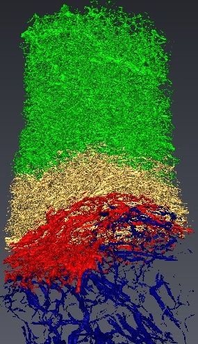 |
Vascular
network segmentation from X-ray micro-tomography : Necrosis (blue),
Tumor (red), Tumor Periphery (yellow) and Sane Tissue (green) (joint work with IMFT, CerCo and ESRF). |
|
|
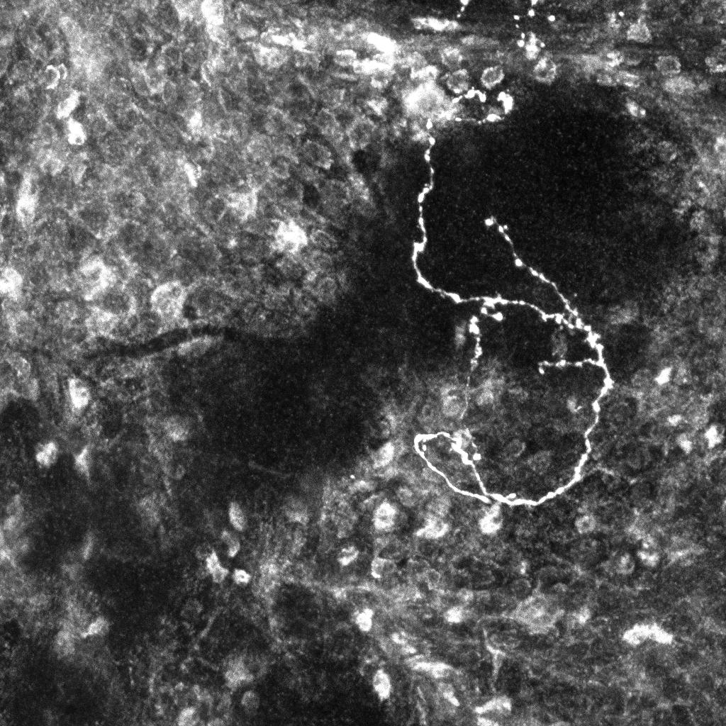
Axonal tree of a labeled
mature neuron. This static image has been acquired from a normal whole
mount brain using a fluorescent confocal microscope (ZEISS LSM510 META
available at the PRISM IBDC imaging platform). Our objective is to
automatically extract the axonal tree.
|
 |
|
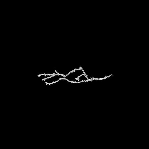 |
3D extracted axon from a static confocal microscopy image. | |
|
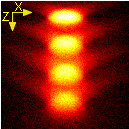
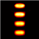
Slice of a simulated
image of a wide field microscope of 4 spherical beads (top) and
restored image using a space-variant restoration approach (bottom) (http://hal.inria.fr/inria-00602650/fr/)
|
  |