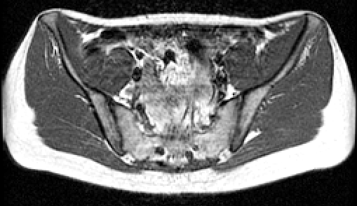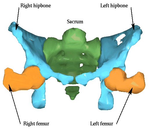

A slice of one of the images. Bones are delimited by a thin border of weak signal that can diseappear in some places.
Image acquisition: Dr. O. Dourthe
Images are MRI T1 weighted, which means that bones are not well visible. Moreover, there is an anisotropy of a factor 4.5 in the vertical direction, which disables the use of most automatique segmentation tools. We have thus used hand segmentations performed by a specialist of MRI.

Segmentation: Dr. O. Dourthe