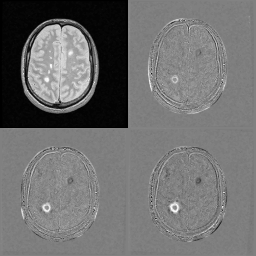
We use here images from an extensive study of multiple sclerosis evolution performed by Dr. R. Kikinis at the Brigham and Women's Hospital (Boston). Each patient typically has 24 MRI in a year. The goal is to register precisely all the images to perform quantitative measurements on the multiple sclerosis evolution.

The same slice in 4 consecutive images after registration. To stress the evolution, the first slice is subtracted from the others. We can see an increasing lesion in white on the left posterior hemisphere and a regressing one in black on the right anterior hemisphere.
Images courtesy of R. Kikinis, processing of J.P. Thirion.