Image at time 1 (I1):
-
The images of this demo are from the Brigham and Women's Hospital (Dr
Charles
Guttman and Dr Ron Kikinis) and some results come from the data
of the EC-funded BIOMORPH
Project.
-
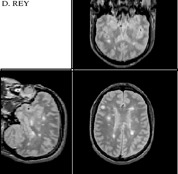
Images at times 1 and 2 consist in 256*256*54 images with voxel size of
0.9*0.9*3.0. There is about 1 month between time 1 and time 2. The images
are MRI, T2 weighted.
-
Our main experiments have been on Multiple Sclerosis disease evolution
over time, but the algorithms are generic and should work for other processes
(evolution of tumors, motion of anatomical structures, ...)
Back to the diagram