Shape reconstruction in medical imaging

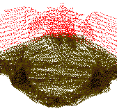
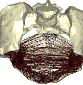
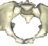
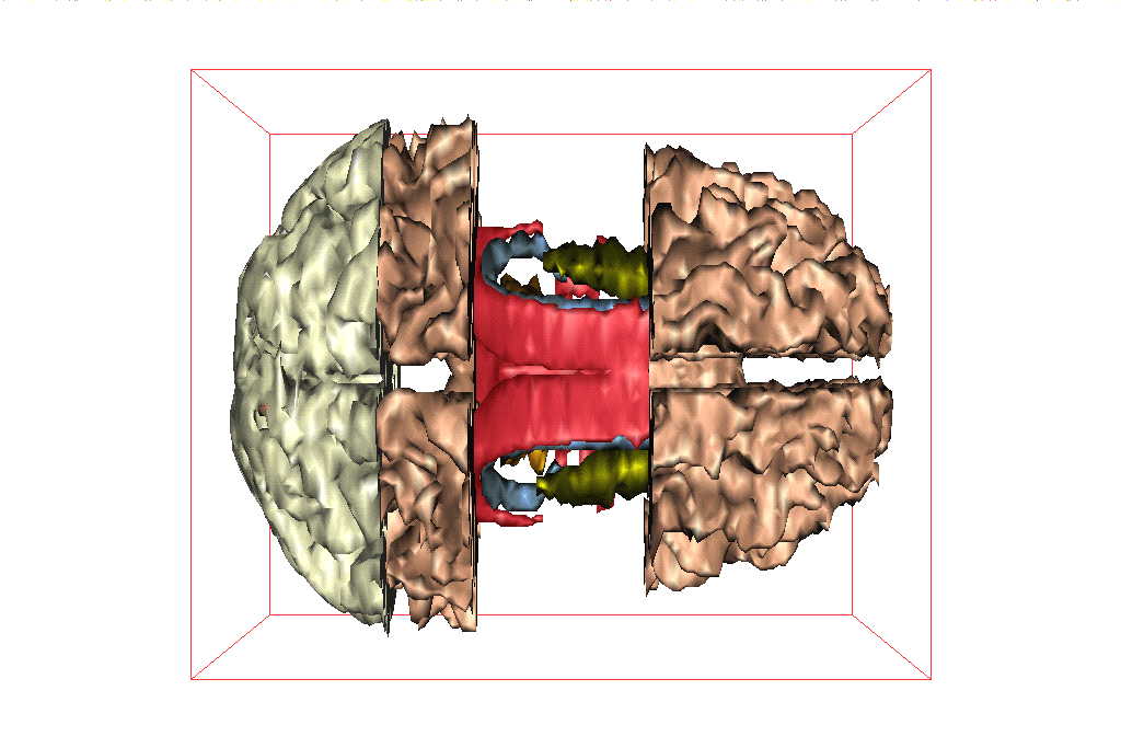
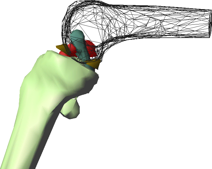
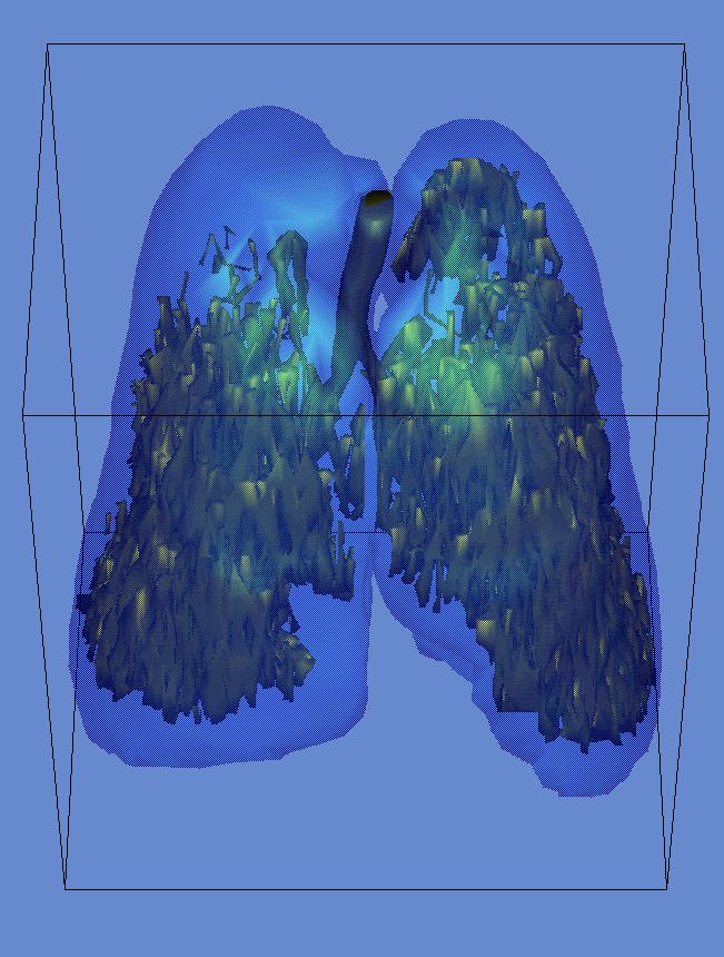
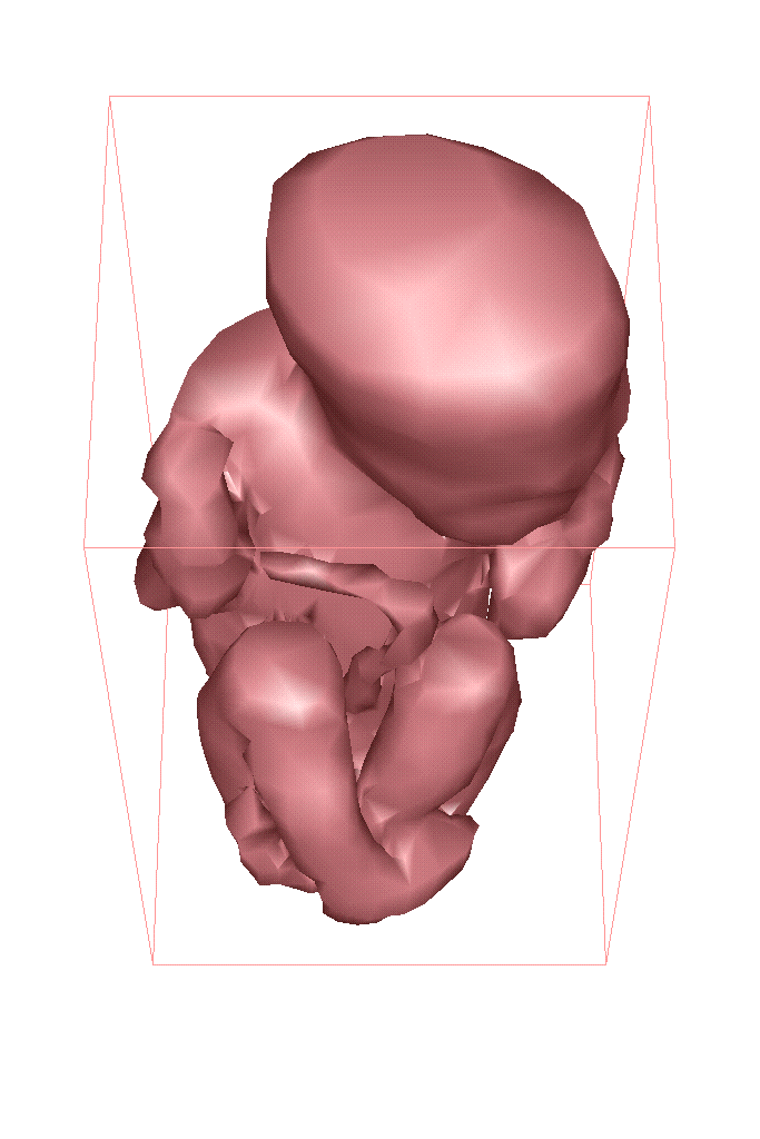
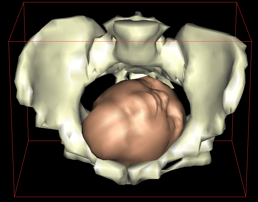
Reconstruction from planar parallel cross sections

The data are generally provided by a medical system such as magnetic resonance (MR) which gives several parallel cross-sections of the desired zone. One can then extract the polygonal contours in every cross section, either manually or using tools from image analysis.

The reconstruction method uses the 3D Delaunay triangulations between every pair of consecutive cross section. In the worst case, this triangulation could be quadratic, but in practise it is quasi-linear. The computation time is output-sensitive. Then all the tetrahedra that can be determined as being outside are removed, and what remains is a tetrahedralization of the reconstructed object.

From left to right, one may observe the Delaunay triangulations, the removal of the outside tetrahedra, and the smoothed reconstructed object.

This reconstruction is volumic. So, it allows simulations using the graph of the Delaunay tetrahedra.
The NUAGES software and a data base

Examples of reconstruction

Click on a picture to view it in real-size
Skull (description) Trachea (description) Brain (description)
Knee-joint (description) Emphysema (description) Fetus (description)

Simulation

Childbirth delivery (description) Knee Prosthesis (description) Endoscopy (description)


Last modified: Fri Jan 21 14:58:44 MET 2000 Monique Teillaud Topics Accueil PRISME Français