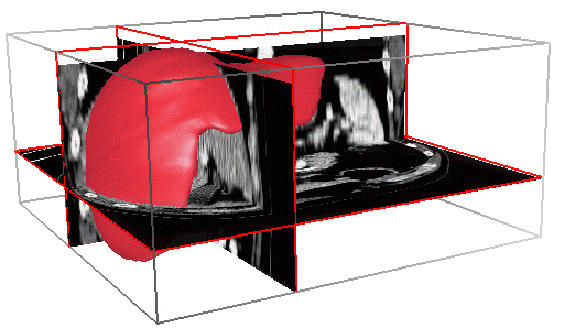
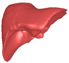
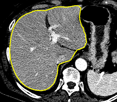
Deformable models are often use for segmentation. We illustrate the
segmentation of the liver in abdominal CT-scan images. A
geometric model of the liver is initialize inside the
volumetric image and deformed toward image edges.
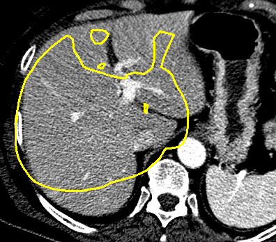
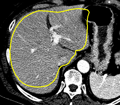
The segmentation process requires different stages:
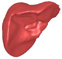
|
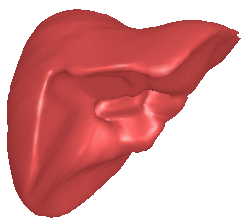
|
| (a) Initialization | (b) Rigid and affine registration |
|
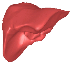
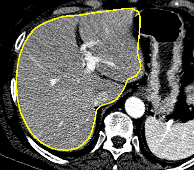
|
| (c) Hybrid deformations (l low) | (d) Hybrid deformations (l high) |
The following figure illusrtates the evolution of a model cut on one slice of the 3D image.
|
|
| (a) | (b) |
|
|
| (c) | (d) |