Epidaure Research Project INRIA Sophia Antipolis


An ElectroMechanical Model Of the Heart for Cardiac Image Analysis

Here are some graphical results of the model described in the publications, in image, video and VRML mesh formats. You can also find additional information on the web pages of the Collaborative Research Action ICEMA (mainly in french) and ICEMA2.
Images:

(click on images to enlarge)

Simulated Electrical Propagation Isochrones:

electrical isochrones electrical isochrones electrical isochrones electrical isochrones

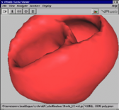
Simulated Electromechanical Contraction and visualisation of the result on the left ventricle endocardium:

electromechanical contraction electromechanical contraction electromechanical contraction electromechanical contraction
Left Ventricle Contraction

3D Image Segmentation with a volumetric deformable biomechanical model:

biomechanical
      model in image biomechanical
      model in image biomechanical
      model in image biomechanical
      model in image

Electromechanical Model in a 3D Image

model in
      echocardiography model in
      echocardiography model in
      echocardiography

Videos:

Electrical wave propagation

Electrical wave propagation + Mechanical contraction

Interaction with 3D images

Between the canine heart model and a segmented image of the Visible Man

Between the canine heart model and a human cardiac MRI (rigid -> similarity -> affine -> local deformation)

Meshes:

beating heart VRML
	   meshes Compressed VRML1 files of the beating heart mesh during the simulated cycle (or compressed archive of all the files). Each mesh represents a 0.01 s step of the simulation. These meshes are the volumetric electromechanical model extracted surfaces.
Original myocardium geometry and fiber directions are courtesy of the Cardiac Mechanics Research Group of UCSD.
Copyright 2002 © Epidaure Research Project - All Rights Reserved - ()