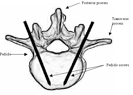
Figure 1.1 Entry point and orientation of transpedicle screws
Robots have had a significant impact on the manufacturing environment, yielding higher productivity and improved quality. Robots, however, have not yet made a significant impact in health care. The application of robots in medical practice is limited by a lack of tools yet to be developed. These tools are different from those in the manufacturing environment. Better bone-surface identification methods, visualization, and control of the manipulator, as well as pre-operative simulations, are of great interest to a surgeon in a robotic-assisted medical intervention. Identification of bone geometry with respect to the manipulator is called registration. (Abdel-Malek et al. 1997)
The proposed thesis is aimed at developing such an identification method.
In spine surgery, spinal instrumentation is often used to apply forces to correct deformity or to stabilize an injured spine. Spine instrumentation may also improve the rate of biologic fusion. In most of this instrumentation, pedicle screws have to be inserted into known position and orientation determined by CT scans. One of the most critical issues to success is the exact insertion of pedicle screws. However, knowledge of the ideal positioning is one thing and accurately achieving it physically is quite another. The space where each screw can be inserted is extremely limited and is close to vital mechanisms and nerves.

Figure 1.1 Entry point and orientation of transpedicle screws
Drill holes and screw placement by human hands have limited accuracy even in the hands of the most gifted surgeon under ideal conditions. Placement of screws in the vertebral pedicle demands the greatest accuracy of screw placement; perhaps greater than is consistently and humanly possible. However, based upon recent work by Abdel-Malek et al. (1997), it has been established that robot-assisted insertion of pedicle screws is a challenging, yet achievable problem. This thesis research aimed at providing a method of registration that accepts CT scans and digitized camera pictures as input, and will produce an appropriate coordinate system identifying the bone geometry. It is believed that the proposed registration system will allow a robotic arm to accurately identify any point on or within a vertebral body. To do this, the relative 3D coordinates of the bone must be known through a proposed stereo vision with the steps:
To develop a matching algorithm between 3D-shape data and actual CT model
To register the bone surface with respect to the robot frame
To develop a bone-identification scheme whereby a surgeon can locate points and directions of pedicle screw insertion